Take your health into your own hands

Take your health into your own hands

Take your health into your own hands

Solar time: here’s why it’s good for our brains

For many people, the solar time brings with it many negative aspects, related to the fact that the days become shorter and the darkness of night overtakes more quickly, giving us the impression that we do not have enough time to carry out our regular daily chores. Actually, this is not quite the case; in fact, in some respects it is quite the opposite. In fact, giving up an hour of evening glow in exchange for an hour of full morning light has so many beneficial effects on our bodies, starting right from our brains.

And it is precisely these positive aspects that need to be focused on, especially for those individuals who are negatively affected by the transition from the summer months (associated with heat, vacations, and relaxation) to the winter months (when we return to the routine of school and work and temperatures drop precipitously). As an article in The Conversation magazine reports, an extra hour of sunshine in the morning brings great benefit to our brains, our moods and makes for a much more productive day. Let’s see why!

Nature and autonomous clock

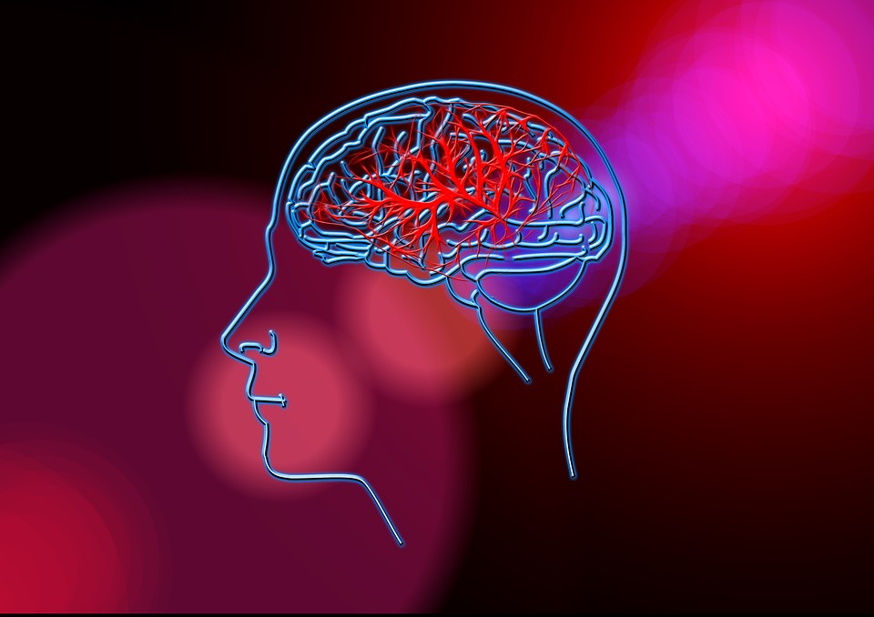
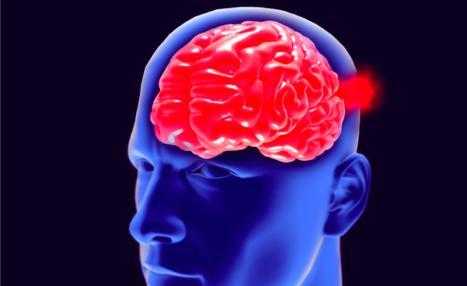
All life on our planet is marked by the alternation of day and night during the 24-hour period. These two “moments” of the day mark our rhythms and influence our biological functions beyond what can be perceived by sight. For this reason, in fact, we become sleepy as evening approaches, and it is more difficult to wake up if we are in an environment that is still dark. In fact, the intensity of light is perceived by special cells in the retina that are directly connected with the suprachiasmatic nucleus, a group of neurons that is responsible for regulating the circadian rhythms ( the changes in biological activities ) of our body. And this is where what we most commonly call the “biological clock” is located.

Get off to a flying start thanks to the sun

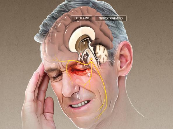
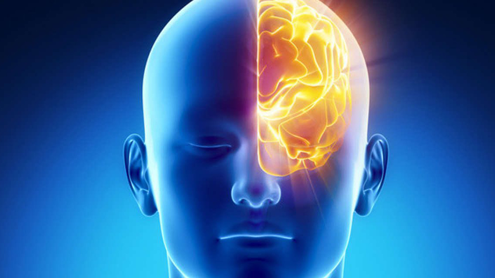
This whole mechanism allows our brain to regulate the amount of hormones to be produced based on the amount of light around us. About 30 minutes after morning awakening, there is a major release of cortisol (called cortisol awakening response, CAR). This hormone is critical for starting the day with the right drive and energy needed. Higher amounts of cortisol in fact have been associated with higher learning capacity and better brain plasticity, as well as a more pronounced ability to make decisions and plan. The release of this hormone occurs more strongly when we wake up in an environment filled with natural light, and its beneficial effects on the body are greater.

For this reason, being able to have an extra hour of sunlight in the morning only gives us a greater charge to start our daily routine in the best and most hassle-free way possible.

Emotions set teenage brains in motion

It is not difficult to think that a particularly strong emotional state can cause increased brain activity, especially in adolescents. The finding is interesting especially when compared to other mechanisms that put our brains to work, mechanisms, however, that require less participation than emotion processing. This issue has been the focus of a very interesting research study resulting from a collaboration between IRCCS Medea and the Bosisio Parini Pole of the same institute, with the IRCCS Ca’ Granda Ospedale Maggiore Policlinico Foundation and the University of Milan, published in the journal Brain and Cognition.

The experiment

The research subjects were a group of boys between the ages of 14 and 19. They were asked to mentally read, in two distinct stages, first emotional verbs and then verbs describing actions. In both cases the subjects had to imagine themselves in the corresponding situations. From the study of MRIs performed on the boys who took part in the experiment, it was found that interaction with verbs describing emotional situations causes increased activation in two precise areas of the brain.

According to the project manager, Barbara Tomasino, the areas of the brain that are most “activated” during this phase are those where, according to the expert’s words, “emotional awareness related to parts of our body is encoded, such as when sweating increases due to a state of anxiety or the heart rate speeds up due to fear.” This whole mechanism is not activated when the brain is busy decoding verbs that describe actions or an emotional situation but with a cognitive task, such as identifying a specific letter within the verb. In fact, as Barbara Tomasino also states, “Therefore, it is not enough to think of the verb to love for the brain areas involved in decoding this emotional experience to be activated, but the feelings corresponding to love must also be imagined.”

This particular brain activity, which is strongly present in boys, also occurs in adult brains, but with less intensity. Finally, according to Paolo Brambilla of the University of Milan, this very interesting study “paves the way toward investigating those emotional situations often found in psychopathology in children and adults suffering from anxiety, phobias or depression.”

A sedentary lifestyle negatively affects memory

Sedentary Lifestyle: It is known that an excessively sedentary lifestyle is strongly detrimental to the health and well-being of our bodies. Spending too many hours a day sitting in a chair behind a desk (as in the case of office or study work) or, even worse, spending entire days on the couch watching TV (perhaps accompanied by unhealthy snacks) Are habits that strongly and negatively affect our health. Among the most prevalent risks are definitely problems related to the health of our heart, which is more prone to dangerous diseases in overly “armchair-loving” individuals. But the list is long, and in addition to the aforementioned cardiovascular problems, it also includes diabetes and the possibility of premature death more generally.

As if that were not enough, according to a recent study, an overly sedentary lifestyle would also have negative effects on our brains, particularly on the section that handles memory.

The study

According to a study conducted by researchers at theUniversity of California, Los Angeles, involving about 35 people between the ages of 45 and 75, a sedentary lifestyle would lead to significant changes in the area of the brain that is critical for memory. All research participants first underwent a cognitive test to find out their daily habits and routines, with specific questions about their (possible) physical activity and the average number of hours spent sitting during the day. The questions were more concerned with the period including the week before the test.

After the test, all participants underwent a high-resolution MRI to get a clear look at the medial temporal lobe, the portion of the brain that is actively involved in the formation of new memories. From the examinations, it could be seen that a very sedentary lifestyle leads to thinning of the medial temporal section of the brain. Such a situation can lead to the decline of an individual’s cognitive abilities and the onset of dementia as the years progress.

How to remedy

The study also brought out that physical activity alone is not enough to improve this condition and protect our brain health. Therefore, it is necessary to change one’s habits and lifestyle. One should avoid spending too many consecutive hours of the day sitting in a chair, but it would be advisable, from time to time during the course of the day, to take short walks, even of a few dozen minutes (just think that to make up for an entire day spent in a chair one needs to take at least an hour of walking). Those most at risk are those who perform office work, but again, you should take advantage of any time of day when you have a few minutes off to do small physical exercises to stretch your arms and legs. It is also helpful to attend events that promote a healthy lifestyle to learn useful tips on how to move without disrupting one’s usual routine.

The brains of abandoned children

Alterations in key nerve circuits, but with family foster care, recovery is possible

A research team from Harvard Medical School‘s Boston Children’s Hospital has documented, in JAMA Pediatrics, the alterations that occur in the brain circuits of children who lived in orphanages in Bucharest. The study, which began in 2001, is part of a project, called the Bucharest Early Intervention Project, involving the 6 orphanages in the Romanian capital, three U.S. Universities led by Harvard, three Foundations, led by the Mac Arthur Foundation of Chicago, whose goal is to examine the effects of childhood institutionalization on brain and behavioral development and, at the same time, to test whether family foster care has the ability to recover from brain damage.

The characteristics of the study are very unique. 136 children around two years of age, who had been in an orphanage since birth or at least for a few months after birth, were randomly divided (randomized) into two groups, one sent to foster care and the other who remained in the orphanage. A choice that instantly puzzles, but it should be known that, prior to this project, there was no adoption program in Bucharest, which therefore begins with the activity of the U.S. group, which covers the costs of fostering and the training of social workers who will be responsible for providing ongoing support to foster families. Finally, the study included a control group consisting of Bucharest children of the same age living with their families. All children were observed for about 8 years at regular intervals, monitoring their intellectual and behavioral development until then ages 9 to 11. Finally, a sample from each of the three groups was selected to be subjected to a meticulous and extensive brain investigation carried out using the Diffusion Tensor technique. This technique, abbreviated DTI (tensor diffusion imaging), allows visualization of the bundles of white matter fibers that connect brain areas to each other.

Children in orphanages showed alterations in white matter microstructure in a number of circuits and namely: the central part (so-called body) of the corpus callosum, cingulum, corona radiata, fornix, external capsule, retro-lenticular area of the internal capsule, and medial lemniscus. Brain images of all these brain circuits of institutionalized children, with the only exception of the retro-lenticular area of the internal capsule, show linkage deficits, which also explain the behavioral, cognitive and emotion management disorders that, with greater frequency, are present in these abandoned children. Intriguing is the exception of the retro-lenticular area of the inner capsule, which, instead of ‘weakening, is shown to be thickened and thus more functional. This area is part of the visual system and is the sensory area studied along with the medial lemniscus, which is a nerve pathway that transports sensation from the body to the brain, which, on the other hand, is deficient like all other circuits. One would think that in institutionalized orphans there would be greater visual acuity, as a need to be alert at all times, and poor tactile sensitivity, a dulling of the senses from lack of caresses and more generally from poor human contact .

Children in foster care, on the other hand, show brain images quite similar to children living with families, although some alterations in the white matter are still visible in the corpus callosum and corona radiata. This study is of great importance not because it is the first. Most recently, work by RJ Davidson’s group at the University of Wisconsin-Madison, in Biological Psychiatry on Feb. 15, documents, in children who have been abandoned or subjected to physical abuse or economic misery, structural alterations on key gray matter areas such as the amygdala and hippocampus. The importance of the Harvard study lies in the fact that it is a randomized controlled, prospective design with within it the demonstration that family foster care is not only an ethical obligation to these “innocents” (after whom the famous orphanage in Florence was named), but it is also an effective health intervention, which even in our country, which has abolished orphanages since 2006, it would be appropriate to extend without delay. There are about 15,000 minors in foster homes, which are certainly not orphanages but are obviously not families either, with an estimated economic cost of more than 40,000 euros per year per child. This money could be better spent.

by Francesco Bottaccioli

Physical and cognitive activity protect women’s brains

Any physical and cognitiveactivity performed by women around age 40-55 can protect intellectual function in later life years, reducing the risk of developing cognitive decline and dementia of various types and levels of severity. Had you already been told this? Most likely, yes, because there have been many studies in this area conducted in recent decades.

The added value inherent in the results of the research conducted at the University of Gothenburg (Sweden) is the particularly long observation period (follow-up was as long as 44 years) and the clarification of the protective effects of physical and cognitive activity against vascular dementia and Alzheimer’s disease, respectively.

The study, recently published in the peer-reviewed journal Neurology, involved 800 women with a mean age at enrollment of 44 years (in the range of 38-54 years), selected from the general population. For all participants, intellectual, artistic, manual, social, religious and sports activities habitually performed were recorded at the beginning of the evaluation, and periodic monitoring of cognitive performance was, therefore, initiated from 1968 to 2012.

During follow-up, 194 women were diagnosed with dementia, 102 with Alzheimer’s disease, 27 with vascular dementia, 41 with mixed dementia, and 81 with dementia associated with cerebrovascular disease. All diagnoses were made based on the criteria provided in the reporting period, taking into account the outcomes of neuropsychiatric interviews, visits and examinations, data collected in medical records and patient registries.

Analysis of available information showed that, in general, women who remain intellectually active between the ages of 40 and 55 have a reduced risk of developing a form of dementia by about one-third (-34%). The protection was found to be particularly pronounced against Alzheimer’s disease, the occurrence of which would be practically halved (-46%) compared to women who were less inclined to read, study, listen to music, take classes, attend museums, go to the theater or movies.

Even more favorable has been shown to be the impact o fexercise, particularly against mixed dementia and cerebrovascular disease-related forms. In fact, the risk of developing the former was found to be 57 percent lower among the more physically active women, while the risk of developing dementia related to cerebrovascular disease was found to be 53 percent lower.

The magnitude of these preventive effects is surprising, to say the least, when one considers that, at present, medical has very few weapons (moreover, of very limited effectiveness) to protect against these neurodegenerative diseases, and that the outcomes cited were obtained after excluding major confounding factors such as educational level, socioeconomic status, presence of hypertension, diabetes or cardiovascular disease, body weight, smoking, stress and depression.

Of course, more confirmation is needed before we can come to any firm conclusions, but in the meantime, it’s worth banishing all laziness and trying to keep as intellectually and physically active as possible, shall we?

Source

Najar J et al. Cognitive and physical activity and dementia. A 44-year longitudinal population study of women. Neurology 2019;92:e1322-e1330. doi:10.1212/WNL.0000000000007021 (https://n.neurology.org/content/92/12/e1322.long)

Female brain ages less than male brain

The aging of the human brain varies greatly from subject to subject: some individuals experience rapid cognitive decline, while others maintain their cognitive abilities intact even in old age. According to some studies, brain aging would be linked primarily to impaired brain metabolism, particularly glucose metabolism, and not only to neurodegenerative phenomena, which is the currently most accepted thesis.

In this line of research is a recent paper, published in a prestigious American journal, which aimed to show that sex differences are able to influence the morphology and physiology of the brain during development and aging.

Through the use of data acquired by brain PET scan from 205 normal adult subjects, aged 20 to 82 years, it was found that throughout adult life the female brain, compared with the male brain, has a persistently lower metabolic brain age than the chronological brain age.

According to the authors of the article, belonging to the Washington University School of Medicine. St. Louis, sexual differences would be able to influence brain aging, in favor of women, the whose brains, with the same age of birth, would be a few years younger than The male one.

SOURCE

Manu S. Goyal et al. Persistent metabolic youth in the aging female brain. PNAS February 19, 2019 116 (8) 3251-3255.

Stroke: here’s what to do

Stroke is a life-threatening emergency. It occurs when a blood vessel in the brain ruptures or becomes obstructed, due to a blood clot. In this case, the brain does not receive the blood flow it needs, and without oxygen, brain cells do not function and die within minutes.

Warning signs that indicate a possible stroke include numbness, weakness or paralysis of one side of the body, blurred vision, difficulty speaking, loss of balance, and sudden headache. Ten percent of strokes are preceded by a transient ischemic attack, the symptoms of which are brief stroke-like episodes. The average transient ischemic attack lasts 1 minute, although some even last for hours. Unlike stroke, the victim returns to normal at the end of the ischemic episode. However, it is important not to ignore the symptoms and take the patient to the hospital.

What do?

  1. Check vital signs.
  2. Place the victim in an appropriate position, if conscious raise the head and shoulders. If, on the other hand, the victim is unconscious put her on her side.
  3. Do not give anything to drink or eat.
  4. Call the ambulance.

Source: Mediserve‘s Pocket Guide to First Aid.

Closed head trauma

In the case of closed head injury, loss of consciousness may not be immediate; in fact, several minutes may elapse between the time of injury and the appearance of neurological signs.

To determine the state of consciousness, the Glasgow Coma Scale, through which a score of 8 or below determines severe trauma.

It is necessary to provide the patient with controlled ventilation and transfer him to intensive care unit.

Source: Mediserve‘s Medical Emergencies in Pediatrics.

What damages the brain (and how to protect it)

Directing center of every physical and psychic function, the Brain is the most complex and delicate organ in the entire human body. For this is well enclosed and protected by the skull bones and partially isolated from what takes place in the rest of the body by the blood-brain barrier. Despite these two anatomical-functional arrangements, there are some number of factors internal and external to the body that can harm it, in Acute or long-distance mode. Here are what are the main enemies of the brain e Some tips to protect it.

Insomnia, identified areas of the brain at risk

Insomnia is widespread, globally, in fact about 770 million people are affected by the problem.

However, still little is known about the factors that may cause difficulty in falling asleep.

A group of researchers from multiple nations, led by international research scientists from the Vrije Universiteit in Amsterdam, has developed and published a study in the pages of Nature Genetics that focuses on those brain processes that affect the risk of insomnia.

The disorder

Insomnia can be defined as a frequent and persistent disorder in which the affected person faces great difficulty in falling asleep and suffers frequent awakenings, which are followed by other difficulties in falling asleep again, even when there seems to be no environmental impediment.

If these complaints persist over time and last for months , surely we can speak of chronic insomnia.

Science

To this day, scientific research has not yet been able to identify which genes may be responsible and their negative effects on the brain.

In order to shed light in research Danielle Posthuma of the Vrije Universiteit and Eus Van Someren of the Netherlands Institute of Neuroscience have assembled a team of scientists, who for the first time have been able to put together so much data and information about the dna and specific sleep habits of a vast number of people, well 1.3 million. They used information and resources from the Uk Biobank and 23andMe database, thereby creating the largest and most important “genetic dataset” to date ever available.

The genes identified

The researchers were able to identify, based on the processing of the vast amount of observed data, the remarkable number of 956 genes, which according to different variants could account for contributions to an increased risk of insomnia.

Then the researchers focused their studies to understand what biological processes, what types of cells and what areas of the brain from the genes used.

It was determined that some of the genes studied play an important role in the functionality of axons, those extensions of neurons that participate in the exchange of information between nerve cells.

Scientists say other genes are found to be active in specific cells, the medium spiny neurons, which are present in the frontal cortex and subcortical nuclei, those areas of the brain known to influence sleep quality.

Mens sana in corpore sano!

Even the Ancient Romans were aware of the positive correlation between exercise and mental activity. But not all sports are the same.

Which sports promote mental activity and what is the optimal amount of physical activity?

These questions were attempted to be answered by a group of researchers from the University of Basel in Switzerland and the University of Tsukuba in Japan in a study published in the journal Nature Human Behaviour.

A sport that includes training based on the development of strength and endurance seems to be more effective in terms of its effects on cognitive performance. Even more positive effects come from those sports activities that require complex coordination patterns and interaction with other players.

However, cognitive improvement does not seem to correlate with higher training intensity, but rather with longer training duration.

The benefits affect all age groups, potentially especially childhood, then the stage of greatest cognitive development, and old age, which is the stage of degradation. Based on the research results, however, it was not possible to define an indicator of the effectiveness of different sports activities according to different age groups. Instead, it has been seen that different ages can be brought together in the same sporting activity, such as through existing experiences of grandparents and grandchildren exercising together or men and boys engaging in more intense sporting activities.

We already know that the volume of sports activity is more important for men’s physical fitness than for women’s. This also applies to mental fitness. Especially in reference to the intensity of movement. While men and boys enjoy significant improvement, and over a longer period, in cognitive performance following hard training and increased, albeit gradual, intensity, for women the improvement in cognitive ability appears to be linked with low to medium intensity sports activities.

Brain aneurysm: what is it?

Cerebral aneurysm is a permanent protuberance, that is, a focal dilatation of an arterial vessel in the brain at which the wall stretches, thinning and forming a bubble. It can result in rupture of the wall at the point where the vessel is most fragile or thinned, causing hemorrhage. Blood loss in the brain can be a cause of hemorrhagic stroke.

Read more.

Anorexia nervosa

Anorexia nervosa was included in DSM 5 (2013) in the broader diagnostic category called Nutrition and Eating Disorders.

The main characteristic of this disorder is food refusal. The term anorexia (literally lack of appetite) is incorrect to describe a disorder in which appetite is in most cases preserved. Rather what characterizes the anorexic person is the terror of gaining weight and the need to control eating in pursuit of thinness.

Read more.

Stroke: ischemic and hemorrhagic

Stroke is a serious disease that occurs as a result of the reduction, or interruption, of blood flow to the brain (by which we mean the brain, cerebellum, and brainstem enclosed in the brainbox) and the subsequent death of brain cells.

Two forms are distinguished: the most common, ischemic stroke, occurs when a blood clot obstructs an artery carrying blood to the brain; hemorrhagic stroke is caused by the rupture of an artery.

A transient ischemic attack (TIA) is when the blood supply to the brain is interrupted for a short time.

Read more.

Morton’s neuroma

Morton’s Neuroma is a neuropathy consisting of a swelling of a nerve in the foot located between the third and fourth toes. The condition is named after the American surgeon Thomas George Morton, who first diagnosed it in the 1800s. Neuroma, with the passage of time, can lead to the development of fibrous scar tissue around the nerve affected by the disorder because of the continuous friction of the adjacent metatarsal bones and deep intermetatarsal ligament, which, at the level of the third space, are more mobile than in other parts of the foot. Although the condition can occur at all ages, it mostly affects women between 40 and 50 years old.

Read more.

Deep Brain Stimulation (DBS)

Deep brain stimulation (DBS from the English acronym Deep Brain Stimulation) is a surgical treatment with the goal of reducing debilitating motor symptoms that characterize movement disorders such as Parkinson’s, dystonia, and essential tremor. It is also a procedure used to treat epilepsy, chronic pain, and obsessive-compulsive disorders.

Read more.

OSAS – Obstructive Sleep Apnea Syndrome

From the English Obstructive Sleep Apnoea Syndrome or.
Obstructive Sleep Apnea Syndrome.
. This is a condition related to repetitive partial or complete obstruction of the first airway at the level of the throat, which is largely underdiagnosed due to both little information about it and confusing symptomatology. It is usually manifested by daytime sleepiness that originates from disturbed sleep due to obstructed airways, causing interrupted breathing. It falls within the macro-area of sleep disorders, more correctly known as “Sleep Disorders.”

Read more.

Head trauma: causes and treatment

Head injury is, in general, damage to the cranio-brain district caused by any physical event of a mechanical nature. Head trauma is responsible for 50% of all traumatic deaths and 2% of all deaths.

In industrialized societies, the major cause of head injuries, in people under 75 years of age, is related to transit accidents (48 percent), in which there is involvement of cars, motorcycles or bicycles and pedestrians. For people over 75 years of age, the majority of head injuries are due to accidental falls and household accidents (25%). Violence is identified as the cause in 4 percent of all head injuries (robbery assault and gunshot wounds, brawling, fighting for trivial reasons), occupational injuries are found in 8 percent, and about 10 percent are due to injuries in the course of sports activities. More than 50% of patients with head trauma have severe associated polytrauma. To adequately understand the multiple injuries that can result, the mechanisms of head injury must first be explained.

Read more.

Brain tumor: what to know

Brain tumors do not have typical symptoms because they induce disorders common to many other diseases of the nervous system. These symptoms depend on the size of the tumor and the part of the brain affected, because each area of the brain governs a particular function, and if that area of the brain is affected the function is impaired.

The most common complaints to people with brain tumors are:

  • Headache
  • Epileptic seizures
  • Memory deficits
  • Depression
  • Cognitive and behavioral disorders
  • Chronic Fatigue (Fatigue)
  • Loss of control of movement or sensitivity to touch
  • Deep venous thrombosis
  • Decreased hearing
  • Decreased vision
  • Endocrine dysfunction

This section describes the most common symptoms of people undergoing treatment for brain tumors, and suggestions on how to deal with them.

Read more.

NUTRITION AND DIET
 
NATURE, SPORTS, PLACES
 
CULTIVATING HEALTH
 
MENOPAUSE
 
MOM IN SHAPE
 
TRADITIONAL CHINESE MEDICINE
 
HEART SURGERY
 
MEDICINES AND MEDICAL DEVICES
 
PARENTING
 
THE CULTURE OF HEALTH
 
HEALTH UTILITIES
 
GENERAL MEDICINE
 
NATURAL MEDICINE, THERMAL
 
MIND AND BRAIN
 
NEUROVEGETATIVE DYSTONIA
 
WAYS OF BEING
 
HEALTH AND SOCIETY
 
HEALTHCARE AND PATIENTS
 
SEXUALITY
 
OLDER AGE
 
CANCERS
 
EMERGENCIES
 
DIGESTIVE SYSTEM
 
RESPIRATORY SYSTEM
 
UROGENITAL SYSTEM
 
HEART AND CIRCULATION
 
SKIN
 
INFECTIOUS DISEASES
 
EYES
 
EARS, NOSE, AND THROAT
 
BONES AND LIGAMENTS
 
ENDOCRINE SYSTEM
 
NERVOUS SYSTEM
 
DIGESTIVE SYSTEM
 
RESPIRATORY SYSTEM
 
UROGENITAL SYSTEM
 
HEART AND CIRCULATION
 
SKIN
 
INFECTIOUS DISEASES
 
EYES
 
EARS, NOSE, AND THROAT
 
BONES AND LIGAMENTS
 
ENDOCRINE SYSTEM
 
NERVOUS SYSTEM
 

your advertising
exclusively ON
MY SPECIAL DOCTOR

complete the form and you will be contacted by one of our managers